Illustration of a nose bleed

Unraveling vWF: A Better Understanding of the von Willebrand Factor's A2 Domain

A team of Lehigh researchers works to characterize blood's mysterious protein.

Story by

Kelly Hochbein

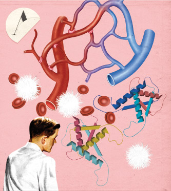
Under normal, healthy circulatory conditions, the von Willebrand Factor (vWF) keeps to itself. The large and mysterious multimeric glycoprotein moves through the blood, balled up tightly, its reaction sites unexposed. But when significant bleeding occurs, it springs into action, initiating the clotting process.

When it works properly, vWF helps stop bleeding and saves lives. However, about one to two percent of the world’s population is affected by vWF mutations that result in bleeding disorders. For those with more rare, severe forms, a very expensive treatment in the form of blood plasma replacement may be required.

On the other hand, if vWF activates where it isn’t needed, it can trigger a stroke or heart attack.

A better understanding of how vWF functions could result in drugs that replace it in those who lack it. It could also lead to the development of new drugs or drug carriers that mimic the protein’s behavior for more effective drug delivery. With that in mind, a team of Lehigh researchers is working to characterize this mysterious protein.

The team, which includes Xuanhong Cheng, associate professor of materials science and engineering; Alparslan Oztekin, professor of mechanical engineering and mechanics; Edmund Webb III, associate professor of mechanical engineering and mechanics; and Frank Zhang, associate professor of bioengineering and mechanical engineering and mechanics; as well as doctoral students Chuqiao Dong, Sagar Kania, Michael Morabito and Yi Wang, is exploring vWF from a variety of angles, through both experiment and simulation.

Illustration of von Willebrand Factor

vWF at Work

At the location of a minor wound, platelets adhere to the collagen-exposed sites near the hole in the blood vessel wall on their own and act as a plug, effectively stopping the bleeding. Rapid blood flow, however, makes it difficult for platelets to do this. Fortunately, the von Willebrand Factor recognizes this rapid blood flow and activates: “It’s a flow-mechanics-activated event, if you will,” explains Webb.

The globular molecule unfolds like a Slinky, stretching to 10 times its original size and exposing its reaction sites. It clings to the broken blood vessel wall, where exposed collagen—the structural protein of the blood vessel wall—attracts platelets. vWF then captures platelets from blood as they flow by, acting like a bridge between the collagen and the platelets.

Although the biological function of vWF has long been recognized by scientists, not much is known about the specifics of how vWF functions, particularly under flow conditions.

“Most proteins in blood functions are executed through biochemical reactions,” says Cheng. “This protein [vWF] also requires some biochemical reaction for its function, so it needs to grab onto platelets, grab onto collagen—those are biochemical reactions. At the same time, vWF relies on mechanical stimulation to execute the biochemical function, and that part is not very well known. That’s what we’re trying to study.”

Adds Webb: “Some of the data that’s coming out of our group but also from other groups indicates that those biochemical reactions are somehow abetted by there being some sort of a tension, a pulling force. So even the biochemical reactions appear to be somewhat mechanically mediated. Again, it was understood that there was this change from a compact, almost ball-like shape, if you will, to this long, stringy thing. But very recently people have been indicating it’s not just that. For this chemical site to be active, you have to be pulling it, you have to be in a bit of tension, locally. So it’s a really fascinating system.”

Hands

Unraveling A2

The von Willebrand Factor is a particularly large protein made up of many monomers, or molecules that can be bonded to other identical molecules to form a polymer. Within each monomer of vWF are different domains: A, C and D. Each domain and each of its respective subdomains has its own role, and many of these roles are yet unknown. The A1 domain, for example, binds vWF to platelets. A3 binds vWF to collagen. The A2 domain unfolds to expose the protein’s reaction sites, and, when fully opened, exposes a site that permits scission of the vWF molecule down to size. Members of the team have focused on the A2 domain, in particular.

“Understanding that domain and how it interacts with the flow, I think, is the best contribution from our group,” says Oztekin.

Each member of the team plays a particular role. Cheng, Zhang and their graduate students work on the experimental side of the project; Oztekin, Webb and their graduate students focus on simulation. Each team’s results inform the work of the other.

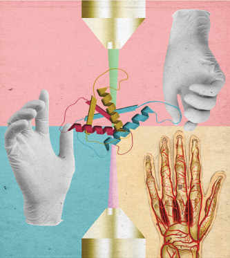
Zhang, who has been studying vWF for years and brought the project to Lehigh, specializes in single-molecule force spectroscopy and mechanosensing, or how cells respond to mechanical stimuli. He uses a specialized tool called optical tweezers, which utilizes a focused laser beam to apply force to objects as small as a single atom.

“Optical tweezers can grab tiny objects,” Zhang explains. “We can grab the vWF and at the same time we apply force to see how the protein changes shape, to see how the proteins are activated when there’s a mechanical perturbation or a mechanical force.”

Cheng develops microfluidic devices, which have a small diameter and can be used to analyze live bioparticles. She and her team make very small channels similar to the geometry of blood vessels—on the order of 10 micron in height, a few millimeters in length and width—so they can mimic the flow condition that vWF encounters in the body. They tag the vWF molecule fluorescently and use a confocal microscope to capture video and still images of the molecule as it flows through the channel at different rates.

“When we talk about this protein under normal flow, it’s one conformation, and then when it’s exposed to certain abnormal flow patterns, you’ll have a different conformation,” Cheng explains. “So we’re trying to characterize or replicate that process in an in vitro system, trying to observe how this protein changes conformation under different flow patterns. And then, if we have mutants versus normal protein, how would they behave differently?”

Doctoral student Yi Wang works with Cheng on the microfluidics channel in which they can observe the vWF molecule unraveling and folding back again in real time under a microscope. To do so, they must create an environment that mimics the shear rate, or change in blood flow velocity, found in the body.

“Because we are using a pretty high shear rate to be comparable to the physiological environment, and because of the limited moving speed of a microscope lens that images the molecule, it’s actually pretty challenging to capture the movement of a molecule if it’s moving,” says Wang.

To solve that problem, the team binds one side of the molecule to the surface of the channel to immobilize it as they apply shear force. They have successfully captured the unfolding phenomenon on video.

“If it [the molecule] is bound too tight, it will just stay there [and not unfold],” says Wang. “If it is too loose, everything will be flushed away. So I was very excited when we got the sweet spot of binding it right there on the surface and so it can unfold and fold back.”

A Better Model

Using data from the experiments, Webb and Oztekin and their doctoral students simulate how vWF behaves or changes conformation in different flow patterns with unprecedented quantitative accuracy. Oztekin is a fluid mechanist who specializes in computational fluid dynamics. He works to understand the blood flow and how vWF reacts to and interacts with it. Webb does atomistic and molecular modeling. The group is using coarse-grain molecular modeling as they seek to answer the question: “How simple can we make this molecular model to get the correct physical behavior that we’re trying to probe but still be able to actually study it computationally?”

Using parameters from experimental results as well as from previous simulations, doctoral student Chuqiao Dong runs explicit atom simulations to get a reasonable model of the A2 domain. Dong’s explicit atom work, says Webb, has inspired the team’s coarse-grain model work.

“[The team is] sort of taking inspiration from Chuqiao [Dong] to then say, ‘Alright, we want to go back to this simpler model, but we want to make it a little less simple based on things we observed in the atomic scale simulations,’” he explains.

Whereas other research teams have utilized a single bead to represent a vWF monomer and its domains, the Lehigh team’s base chain model consists of two beads (the A1 and A3 domains) connected by one spring (the A2 domain) representing one monomer of vWF.

Says doctoral student Michael Morabito: “All of these beads are able to interact with each other through various forces, and they’re also able to interact with each other through the solvent [blood]. So on this level, something that’s very important in our research is understanding what are called hydrodynamic interactions: If one bead moves in the flow field, that displaces the solvent. And when that solvent is displaced, it moves other beads. This one moves, causes this one to move, only through the solvent … The way that all these beads interact is in a very complex nature, which allows us to model these unfolding events and these unraveling events.”

Representing the vWF monomer as one bead as others have previously, says doctoral student Sagar Kania, doesn’t allow for the study of how A2 unfolds because that single bead represents the entire vWF monomer and does not distinguish A2 as a sub-monomer entity. This approach misses “an important piece of the puzzle.”

The Lehigh team, says Kania, is making their model more complicated for more accurate observation of the A2 domain behavior.

“In the old model, they were considering the force [of blood flow] on the bead, but not considering force on the spring [A2],” he says. “The model was such that we couldn’t consider the force on the spring which represents the monomeric entity [A2 domain] in the model. [We now] consider that drag force, or the force by the fluid on the spring that is connected between the beads, to get the critical shear rate.”

The critical shear rate, or the rate of blood flow at which the A2 domain unfolds, is essential to understanding the mechanical properties of vWF. Adopting their model from the experimental data gets the simulation team closer to vWF’s actual architecture, but it then increases the critical shear rate.

“The strength of the blood flow that we had to employ [in our model] in order to make the A2 domain unfold was huge—like orders of magnitude larger than what would really happen in the blood flow,” says Morabito.

Webb explains: “We bring observations like these to our colleagues doing experiments and the ensuing discussions help us better understand the simulation conditions needed to most accurately compare to experiment. In work currently in review, Yi [Wang] and Mike [Morabito] have advanced companion experimental and simulation data that are in remarkable agreement.”

Kania and Morabito are working to maintain in their model the correct molecular architecture of vWF while also incorporating drag on the protein to correctly model its effect.

Says Morabito: “Trying to preserve the architecture of vWF while also correctly capturing its dynamics at the correct blood flow rates has been the most surprising and difficult challenge for me here.”

Inspired Drug Delivery

The team seeks to not only design drugs to replace vWF for those who lack it, but also to utilize vWF’s flow-sensing mechanism to design other types of drugs or drug carriers that can be activated and release drugs in situ when there’s a change in flow pattern.

Zhang and Cheng and their teams are considering whether they can make polymer systems that mimic the behavior of vWF in the in vitro system that can eventually be used in vivo as both a therapeutic drug for bleeding disorders and also as a drug-release carrier. In other words, they hope to engineer polymer systems to mimic the function or behavior of vWF.

“A drug [might be] encapsulated in the polymer until it unfolds, and then the drug is exposed and released at a high-shear stress environment or abnormal flow environment,” she explains. “That’s the general idea.”

In that case, in the event of a stroke, instead of relying on the injection of a critical drug at the hospital, a patient prone to blood clots could already have the drug in his body, dormant until it is activated by changes in the blood-flow environment.

“You can have the drug encapsulated by the carrier and injected,” explains Zhang. “But the carrier, just like the vWF, is inactive—it just protects the drug. When the flow pattern changes, when a stroke happens or the blockade in the vessel happens, this carrier, when it passes by those locations, the drug can be released in situ, right in that location.”

This type of drug delivery might provide an alternative to the risk of circulating a blood-thinning drug through the body of a patient with a bleeding disorder, which could cause problems in areas beyond a blood clot.

“You want to just release drugs where you have the stroke or you have the vessel blockage,” says Zhang.

More knowledge about vWF’s functions can help with other medical problems as well. In certain conditions, vWF can activate where it shouldn’t, creating problems such as thrombosis, or undesired clotting.

“Sometimes in the arteries or in the heart when the patient doesn’t have the heart valve closed completely … vWF is activated by this high shear force and this generates trouble because you don’t want this molecule to be activated,” Zhang explains. “There’s a problem in that scenario. So if we can design drugs or drug carriers mimicking the vWF, it’s a huge advancement.”

Additionally, recent studies have indicated that implanted medical devices can change blood flow conditions and inappropriately activate vWF.

“Flow fields near implants might create enough of the shear forces, as they call it, to open the molecule up and cause thrombosis,” says Webb, “so that can be really bad. There are a lot of unknowns about this, even though there is a lot that’s been learned over the past decade.”

Illustration of head and fire truck

New Discoveries

The team’s collaborative efforts have resulted in several new discoveries, all supporting the notion that the A2 domain is a major player in the mechanical function of vWF. Team members have produced several publications, including a 2018 article in the Biophysical Journal titled “Internal Tensile Force and A2 Domain Unfolding of von Willebrand Factor Multimers,” of which Morabito is the lead author, and another, “Long-Ranged Protein-Glycan Interactions Stabilize von Willebrand Factor A2 Domain from Mechanical Unfolding,” published in Scientific Reports with Dong as the lead author.

“There are no other groups out there that are using this method of simulation to examine intramonomer dynamics,” says Morabito. “We are able to explicitly and deterministically calculate certain properties of interest about the A2 domain without having to make many assumptions or rely on a probabilistic description. Many times in math we’ll have a probability distribution for something to occur, such as A2 domain unfolding, and we’ll go sample this probability distribution to determine if, for example, an A2 domain is unfolded or not at any instant. But we can directly see if it unfolded or not because we’ve explicitly modeled that. So that’s one of the real strengths of our team.”

Working with researchers who conduct experiments provides great benefit, says Dong.

“We can look at one thing from different angles,” she explains. “It’s great because we have [the] experimental [and] we have [the] simulation part, so for our simulation we can get a preview of what it should be like from the experimental data. Also we can get a sense of how large A2 is or what it will look like under different conditions, all the parameters, from experiments.”

The researchers aim to have the computational work and the experimental work continue to move in parallel.

“I can see a single vWF molecule elongating under the microscope, and Mike [Morabito] will have a computational video of a similar size of molecule unraveling,” says Wang. “So we can compare. His work will definitely inspire mine. He will get more parameters more easily than us because it’s kind of harder to control in the experimental side as you have so many variables. And on this side, I think our experimental data can also help him to justify his model and his results, which will be very useful.”

A Dynamic Team

The team is trying to solve a complicated biological puzzle, says Wang—and that makes theirs a very appealing project.

“There are a lot of things going on inside the human body, and we are looking at a very specific one,” she says. “And if you look at the specific one, there are a huge amount of things in this one function. If we can know what exactly is happening, I would think that that is solving a mystery. … It’s already showing us the beauty of nature, the human body functioning.”

Says Morabito: “One thing I really like about this project is that the problem itself necessitates such an interdisciplinary approach. The nature of the problem we’re trying to solve is so complex that there’s no one person able to solve it. … We’re on the cutting edge of it, and that feels good.”

Combining expertise from different disciplines has its challenges, says Webb, but the payoff is valuable—and enjoyable. “There has definitely been a learning curve for our group, a communication curve,” he explains. “I think it’s partly just understanding limitations, capabilities, [and] how do we make this all work and become productive?”

The work has also required researchers to step outside their comfort zones. A team meeting early in the fall 2018 semester featured presentations by the project’s doctoral students, each of whom updated the group on their progress. The group’s energetic and collaborative dynamic had faculty learning from students, students from faculty, faculty from faculty and so on. The excitement in the room was palpable.

“Typically in all of my prior work, the entities we were modeling were so small, or the way that we’re modeling the fluid is such that we didn’t have to worry about representing what are known as the hydrodynamic interactions,” says Webb. “[But] those are incredibly important here. ... [We] are working together to understand, how do we take these molecular models but properly capture those effects? So it’s been really a lot of fun.”

Says Oztekin: “Fun is an understatement. ... Our group is at the peak now. It’s so good.”

This project, “Mechano-Biologically Informed Molecular Models of Flow Sensitive Biopolymers,” is supported by a $1.2 million grant from the National Science Foundation (NSF).

Ebola virus illustration

Zhang and Jagota discovered that the shape and mechanical properties of cells and the [Ebola] virus-like particle are very different.

Understanding Ebola

The rare and deadly Ebola virus has no cure. And at this point, just how the Ebola virus infects its host cell isn’t entirely clear. Frank Zhang and Anand Jagota, the Robert W. Wieseman Chair of Engineering, founding chair of the department of bioengineering and professor of chemical and biomolecular engineering, are trying to find out.

Zhang and Jagota, an expert in computational molecular adhesion mechanics, believe quantitative knowledge about how Ebola interacts with the T-cell immunoglobulin and mucin (TIM) family proteins can help them predict the conditions for Ebola’s attachment to the host cell.

To enter a human cell, Ebola takes advantage of a natural process called macropinocytosis, through which the cell “cleans up” its surroundings by internalizing and eventually destroying the dead cell debris that surrounds it. TIM proteins serve as cell-surface receptors and allow immune cells to recognize the dead cell debris and internalize it. Ebola interacts with the TIM proteins and uses them to hijack this important physiological process, which essentially welcomes the virus into the cell.

Once inside the cell, the virus membrane fuses to the endosome that has formed around it and releases its genetic content into the cell like the proverbial Trojan horse.

“Viral RNA further hijacks the cell mechanism to make proteins,” says Zhang. “At that stage these replicate themselves inside and then they bud off the membrane to form the new virus. Along this process, of course, our cells die and that generates the problem.”

Zhang and Jagota seek to understand how the Ebola virus interacts with the cell membrane and gets internalized so they can eventually help to design a drug that will prevent Ebola from entering the cell while still allowing the natural internalization process of dead cells to take place. Their first step: determining the differences between Ebola and cell debris.

Zhang uses an atomic force microscope (AFM), which scans the surface of a very small sample to determine its mechanical properties: its shape, softness or stiffness. He uses a virus-like particle (VLP), which has the same shape and shell as the Ebola virus but not its genetic materials.

“[We discovered that] the shape and the mechanical properties of cells and VLP are very different,” says Zhang. “We thought maybe we can utilize this difference, and we can hopefully design a drug that would block this stiffer object [the VLP] from coming into the cell but not block the natural process where the cell ingests a soft and round object.”

Zhang also uses the AFM to bring the virus-like particle to the cell with its TIM receptor expressed, observes their interaction, and pulls them apart to determine the mechanical strength of the interaction, or how much force is required to pull them apart.

Jagota uses mathematical models to understand the interaction between Ebola and the cell, what properties represent the Ebola virus—its stiffness, its shape—and what properties represent the cell —the components it presents on its surface—in this interaction.

Illustration by Laurindo Feliciano

This story originally appeared as "Blood's Mysterious First Responder" in the 2019 Lehigh Research Review.

Story by

Kelly Hochbein